Home
/ Shoulder Muscles Diagram : Shoulder Anatomy Diagram With Labels Human Animal Anatomy And Physiology Diagrams Lower Back Anatomy Muscles Neck And Shoulder Muscles Shoulder Muscle Anatomy Muscle Anatomy Troquinhasdabia : Muscles diagram front and back below you'll find several different muscles diagrams.
Shoulder Muscles Diagram : Shoulder Anatomy Diagram With Labels Human Animal Anatomy And Physiology Diagrams Lower Back Anatomy Muscles Neck And Shoulder Muscles Shoulder Muscle Anatomy Muscle Anatomy Troquinhasdabia : Muscles diagram front and back below you'll find several different muscles diagrams.
Shoulder Muscles Diagram : Shoulder Anatomy Diagram With Labels Human Animal Anatomy And Physiology Diagrams Lower Back Anatomy Muscles Neck And Shoulder Muscles Shoulder Muscle Anatomy Muscle Anatomy Troquinhasdabia : Muscles diagram front and back below you'll find several different muscles diagrams.. Other muscles that aid in shoulder movement include: Terms in this set (81). Shoulder joint of human body anatomy infographic diagram with all parts including bones ligaments muscles bursa cavity capsule cartilage membrane for medical science education and health care. Shoulder muscle and ligament diagram. The shoulder muscle tissues can be readily injured and therefore being aware of the appropriate strategy is pretty significant when functioning out.
Ankle muscles diagram, back muscles diagram, chest muscles diagram, diagram of shoulder muscles and tendons, hip muscles diagram, knee muscles diagram, neck muscles diagram, rotator cuff muscles diagram, human muscles. There are three main muscles in your shoulder: The two large main muscles of this. The rotator cuff is a complex and delicate structure of. Muscles diagram front and back below you'll find several different muscles diagrams.
Deltoid Muscle Wikipedia from upload.wikimedia.org Learn faster with interactive shoulder quizzes, diagrams and worksheets. Supraspinatus, infraspinatus, ters minor,.et), using interactive animations and labeled diagrams. Although three ligaments protect and surround the shoulder joint, most of its stability comes from the powerful muscles and tendons of the rotator cuff. This rotator cuff muscle helps with the raising and lowering of the upper arm. Other muscles that aid in shoulder movement include: The clavicle (collarbone), the scapula (shoulder blade), and the humerus (upper arm bone) as well as associated muscles, ligaments and tendons. Major, subscapularis, coracobrachialis anterior/middle deltoid** spine to the humerus pectoralis. Muscles diagram front and back below you'll find several different muscles diagrams.
Terms in this set (81).
The core muscles are those in the abdomen, back, and pelvis, and they. Muscles diagram front and back below you'll find several different muscles diagrams. It is the major joint connecting the upper limb to the trunk. Related online courses on physioplus. The shoulder joint (glenohumeral joint) is a ball and socket joint between the scapula and the humerus. Muscles allow a person to move muscle tendons in the knee joint and the shoulder joint are crucial in stabilization. Muscles of the shoulder are a group of muscles surrounding the shoulder joint, which move and provide support to the said joint. The primary function of the shoulder girdle is to give strength and range of motion to the arm. You can see in the shoulder muscle diagrams that the shoulder is one of the largest and most complex joints in the body. Human anatomy diagrams show internal organs, cells, systems, conditions, symptoms and sickness information and/or tips for healthy living. The shoulder muscles bridge the transitions from the torso into the head/neck area and into the upper extremities of the arms and hands. Tutorials on the shoulder muscles (e.g rotator cuff muscles: This rotator cuff muscle helps with the raising and lowering of the upper arm.
The shoulder joint (glenohumeral joint) is a ball and socket joint between the scapula and the humerus. Printable shoulder muscles diagrams to help you study the muscles structure in human's shoulder. The rotator cuff is a complex and delicate structure of. These muscles aren't as visible as the deltoids, but they are equally (if not more) important. Groin muscles diagram diagram of groin aponeurosis from sscsantry groin project medical.
Shoulder Muscle Diagram Anatomy System Human Body Anatomy Diagram And Chart Images from anatomysystem.com Supraspinatus, infraspinatus, ters minor,.et), using interactive animations and labeled diagrams. See below to view an image of the rotator cuff structure: Related online courses on physioplus. Muscles allow a person to move muscle tendons in the knee joint and the shoulder joint are crucial in stabilization. Diagram shoulder muscles human anatomy shoulder muscles amazing neck and shoulder muscles. Printable shoulder muscles diagrams to help you study the muscles structure in human's shoulder. The two large main muscles of this. This diagram depicts shoulder muscle diagram.
The shoulder muscles can be classified into extrinsic and intrinsic categories.
The shoulder muscles are associated with movements of the upper limb. The shoulder muscle tissues can be readily injured and therefore being aware of the appropriate strategy is pretty significant when functioning out. Diagram muscle shoulder joint (page 1) 2. The extrinsic muscles of the shoulder include trapezius, latissimus dorsi, levator scapulae, rhomboid major and rhomboid minor. Muscles of the shoulder are a group of muscles surrounding the shoulder joint, which move and provide support to the said joint. Just like the muscle tissues in unique elements of the human physique, even our shoulder muscle tissues are prone to standard put on and tear. Ankle muscles diagram, back muscles diagram, chest muscles diagram, diagram of shoulder muscles and tendons, hip muscles diagram, knee muscles diagram, neck muscles diagram, rotator cuff muscles diagram, human muscles. The teres minor, subscapularis, supraspinatus, and infraspinatus muscles together form the rotator cuff, which stabilizes the humeral head (the ball. The muscular system consists of various types of muscle that each play a crucial role in the function of the body. The shoulder muscles produce the characteristic shape of the shoulder and can be classified into two groups: Human anatomy and physiology diagrams: Shoulder muscle and ligament diagram. Learn vocabulary, terms and more with flashcards, games and other study tools.
The shoulder muscle tissues can be readily injured and therefore being aware of the appropriate strategy is pretty significant when functioning out. Other muscles that aid in shoulder movement include: The shoulder muscles can be classified into extrinsic and intrinsic categories. Major, subscapularis, coracobrachialis anterior/middle deltoid** spine to the humerus pectoralis. Diagram muscle shoulder joint (page 1) 2.
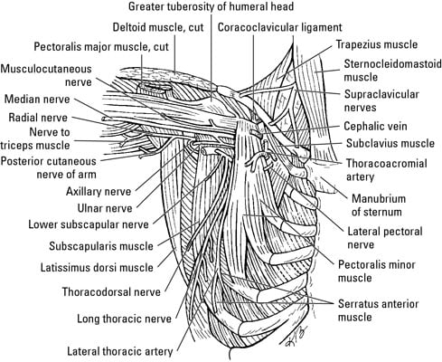
Muscles Of The Shoulder And Arm Dummies from www.dummies.com Major, subscapularis, coracobrachialis anterior/middle deltoid** spine to the humerus pectoralis. Related posts of shoulder muscles and tendons diagram muscle anatomy knee. Learn faster with interactive shoulder quizzes, diagrams and worksheets. Groin muscles diagram diagram of groin aponeurosis from sscsantry groin project medical. The shoulder muscles are associated with movements of the upper limb. There are three main muscles in your shoulder: The shoulder muscles can be classified into extrinsic and intrinsic categories. See below to view an image of the rotator cuff structure:
Shoulder muscle and ligament diagram.
Sternum shoulder muscles **muscles on anterior aspect pec. Terms in this set (81). Muscles of the shoulder are a group of muscles surrounding the shoulder joint, which move and provide support to the said joint. Human anatomy and physiology diagrams: The teres minor, subscapularis, supraspinatus, and infraspinatus muscles together form the rotator cuff, which stabilizes the humeral head (the ball. Although three ligaments protect and surround the shoulder joint, most of its stability comes from the powerful muscles and tendons of the rotator cuff. Shoulder joint of human body anatomy infographic diagram with all parts including bones ligaments muscles bursa cavity capsule cartilage membrane for medical science education and health care. Diagram muscle shoulder joint (page 1) 2. Helps in internal rotation by allowing the individual to rotate the upper arm inwards and in. The primary function of the shoulder girdle is to give strength and range of motion to the arm. Learn vocabulary, terms and more with flashcards, games and other study tools. 6 photos of the shoulder muscles labelled diagram. The shoulder muscles are associated with movements of the upper limb.